
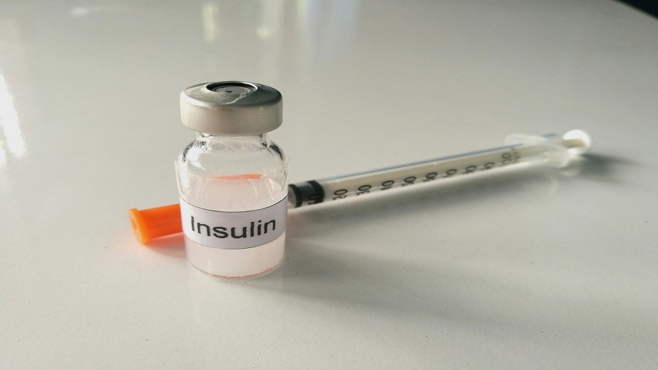
Insulin
⁃ contains an A chain and a B chain, joined by two disulfide bridges.
⁃ Proinsulin is synthesized as a single-chain peptide. Within storage granules, a connecting peptide (C peptide) is removed by proteases to yield insulin. The C peptide is packaged and secreted along with insulin, and its concentration is used to monitor beta cell function in diabetic patients who are receiving exogenous insulin.
1. Regulation of insulin secretion
a. Blood glucose concentration
⁃ is the major factor that regulates insulin secretion.
⁃ Increased blood glucose stimulates insulin secretion. An initial burst of insulin is followed by sustained secretion.
b. Mechanism of insulin secretion
⁃ Glucose, the stimulant for insulin secretion, binds to the Glut 2 receptor on the beta cells.
⁃ Inside the beta cells, glucose is oxidized to ATP, which closes K+ channels in the cell membrane and leads to depolarization of the beta cells. Similar to the action of ATP, sulfonylurea drugs (e.g., tolbutamide, glyburide) stimulate insulin secretion by closing these K+ channels.
⁃ Depolarization opens Ca2+ channels, which leads to an increase in intracellular [Ca2+] and then to secretion of insulin.
2. Insulin receptor
⁃ is found on target tissues for insulin.
⁃ is a tetramer, with two a subunits and two beta subunits.
a. The a subunits are located on the extracellular side of the cell membrane.
b. The Beta subunits span the cell membrane and have intrinsic tyrosine kinase activity. When insulin binds to the receptor, tyrosine kinase is activated and autophosphorylates the J3 subunits. The phosphorylated receptor then phosphorylate& intracellular
proteins.
c. The insulin-receptor complexes enter the target cells.
d. Insulin down-regulates its own receptors in target tissues.
⁃ Therefore, the number of insulin receptors is increased in starvation and decreased in obesity (e.g., type 2 diabetes mellitus).
3. Actions of insulin
⁃ Insulin acts on the liver, adipose tissue, and muscle.
a. Insulin decreases blood glucose concentration by the following mechanisms:
(1) It increases uptake of glucose into target cells by directing the insertion of glucose transporters into cell membranes. As glucose enters the cells, the blood glucose concentration decreases.
(2) It promotes formation of glycogen from glucose in muscle and liver and simultaneously inhibits glycogenolysis.
(3) It decreases gluconeogenesis. Insulin increases the production of fructose 2,6-bisphosphate, increasing phosphofructokinase activity. In effect, substrate is directed away from glucose formation.
b. Insulin decreases blood fatty acid and ketoacid concentrations.
⁃ In adipose tissue, insulin stimulates fat deposition and inhibits lipolysis.
⁃ Insulin inhibits ketoacid formation in the liver because decreased fatty acid degradation provides less acetyl-CoA substrate for ketoacid formation.
c. Insulin decreases blood amino acid concentration.
⁃ Insulin stimulates amino acid uptake into cells, increases protein synthesis, and inhibits protein degradation. Thus, insulin is anabolic.
d. Insulin decreases blood K+ concentration.
⁃ Insulin increases K+ uptake into cells, thereby decreasing blood [K+].
4. Insulin pathophysiology-diabetes mellitus
Case study: A woman is brought to the emergency room. She is hypotensive and breathing rapidly; her breath has the odor of ketones. Analysis of her blood shows severe hyperglycemia, hyperkalemia, and blood gas values that are consistent with
metabolic acidosis.
Explanation:
a. Hyperglycemia
⁃ is consistent with insulin deficiency.
⁃ In the absence of insulin, glucose uptake into cells is decreased, as is storage of glucose as glycogen.
⁃ If tests were performed, the woman's blood would have shown increased levels of both amino acids (because of increased protein catabolism) and fatty acids (because of increased lipolysis).
b. Hypotension
⁃ is a result of ECF volume contraction.
⁃ The high blood glucose concentration results in a high filtered load of glucose that exceeds the reabsorptive capacity (TnJ of the kidney.
⁃ The unreabsorbed glucose acts as an osmotic diuretic in the urine and causes ECF volume contraction.
c. Metabolic acidosis
⁃ is caused by overproduction ofketoacids (~-hydroxybutyrate and acetoacetate).
⁃ The increased ventilation rate, or Kussmaul respiration, is the respiratory compensation for metabolic acidosis.
d. Hyperkalemia
⁃ results from the lack of insulin; normally, insulin promotes K• uptake into cells.